Salamanders can regenerate lost limbs, but mammals cannot. Recent discoveries about cell potency and positional information are helping scientists understand why, while opening new possibilities for regenerative medicine. OUPblog - Academic insights for ...
Everyone knows that many salamanders can regenerate legs and tails while mammals never can (Figs.1, 2). If you tell a layperson that you work on regeneration, they will inevitably ask, “How can they do it when we can’t?” For most of my career, I was obliged to answer, “We don’t know”, and reflect on the fact that the key questions of science really are those simple ones asked by lay people.
The great news of the last few years is that now we do know. At least, now we have some good ideas about how it works, although there are still many gaps to fill. The two key concepts are cell potency and positional information. Cell potency relates to the range of cell types that the cells of a regenerating structure are able to become. Positional information relates to a system of signals that vary with position within the regenerating structure and control what pathway of differentiation those cells adopt.
Fig. 1. The axolotl, a favourite animal for regeneration research. Image credit: Jonathan Slack
Fig. 2. A limb regenerating from an upper arm amputation over 146 days. From Wells, K.M., et al., 2021. eLife 10, e68584.
Let’s start with the cells and consider those cells making up the limb of a vertebrate animal. The skeletal structures and connective tissues of limbs arise in embryonic development from a limb bud—a structure consisting of a loose mass of cells wrapped up in an outer layer. The loose cells are multipotent, meaning that they can become any one of a series of connective tissue cell types: bone, cartilage, tendon, ligament, or dermis (the inner layer of the skin). The other principal structures of a limb: the muscles, nerves and blood vessels, are formed from separate cell populations that grow into the bud during limb development.
When an adult salamander limb is amputated, the connective tissue cells near the cut surface dedifferentiate and become a uniform population of multipotent cells very similar to those of the embryonic limb bud. This fact has only been discovered recently, and it relied on a very powerful modern technology: single-cell RNA sequencing, which enables individual cells to be analysed to find which genes are active and to what extent. This is not the case for the cells of frog tadpole legs, which show very limited regeneration. However, it has been shown that grafting of embryonic limb bud cells into the amputation sites of frogs can improve their regenerative ability. So, to achieve successful regeneration, one requirement is a mechanism to de-differentiate connective tissue cells into multipotent cells similar, or identical to, those of the embryonic limb bud.
But this is only half the story. To regenerate a complex structure like a limb, these cells need to re-differentiate into the various functional cell types arranged in a complex three-dimensional pattern. This is the role of positional information.
Positional information is a concept invented by Lewis Wolpert (1929–2021), a public intellectual well known both in developmental biology and in wider social circles. In the late 1960s, he proposed that developing or regenerating systems must have some kind of embedded information that varies with position, such that multipotent cells can look at the positional information in their locality and use it to decide into what cell type to differentiate. Wolpert postulated the existence of positional signals—for example, gradients of diffusible molecules, which set up the system; and positional values, which were a long-term stable representation of the positional information. This latter concept of positional value is particularly important in regeneration because the information is needed not just for embryonic development, but also to be available to control regeneration after maybe years of the animal’s life.
The identity of many positional signals was discovered between 1980 and 2000 through intensive studies of embryonic development in frogs, mice, fruit flies, and nematode worms. They are composed of small proteins secreted by cells, which are often called “growth factors” or “cytokines” in other contexts. They include molecules discovered by biochemists, like fibroblast growth factors, transforming growth factors, and bone morphogenetic proteins, and others discovered by geneticists with weird names like Wnt and Hedgehog. Most of these factors are active during limb development and again during regeneration. However, they do not persist during adult life. What does persist is the system of positional values, which were only discovered recently.
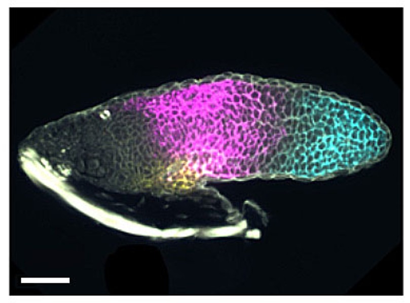
Fig.3. Visualisation of positional values in a larval limb bud. mRNA for shox is magenta, and for hoxa13 is cyan. Scale bar 100µm. From Fig.5 in T.J.Duerr et al. bioRxiv preprint doi: https://doi.org/10.1101/2024.08.07.607055.
The key elements of positional value are the genes that define specific body regions during embryonic development. Such genes all encode transcription factors, which are proteins that control the expression of other genes. For example, in the limb, expression of a gene called shox defines the middle part of the limb and of another gene called hox-a13 defines the hand or foot. These genes are not expressed in the mature limb, but they are still “open for business” in the appropriate regions. For example, hox-a13 is permanently repressed in the upper arm by a specific modification of chromosomal proteins, while in the hand it is not repressed and so it can be re-expressed when exposed to the stimuli of regeneration. In other examples, there is very low-level expression of the relevant positional information genes in the adult, which can be upregulated in regeneration. There are also some cell adhesion molecules, regulated by the same genes, that keep the cell populations in appropriate groups as regeneration proceeds.
Although mammals use the same genes to specify positional information in embryonic development, these genes cannot be reactivated in the adult. Mammals have neither the appropriate multipotent cells nor the appropriate positional information to enable successful regeneration. This is why “they can do it and we can’t.” Whether these key elements can be rebuilt using the modern techniques of regenerative medicine remains a question for the future.
Although the concept of visual abstracts began even before the widespread use of the internet, with journals like Angewandte Chemie and Tetrahedron Lettersfeaturing graphical abstracts in the late 20th century, it wasn’t widely used across all journals until the surge in scholarly papers and information overload. Given that almost 3 million scientific papers were indexed by Scopus and Web of Science alone in 2022, it isn’t surprising that it takes 17 years for research evidence to reach clinical practice. Therefore, for research to influence clinical practice, it must be effectively communicated to key audiences such as policymakers, healthcare professionals, and patients.
Since 2016, Dr Andrew Ibrahim has helped popularize the use of visual abstracts on social media. His studies have shown that presenting research findings in a visual way promises to improve audience reach and drive change, as visual abstracts make use of our ability to rapidly process and retain visual over textual information.
Planning your visual abstract
Paediatrics & Child Healthencourages authors to submit visual or video abstracts with their manuscripts. While visual (or graphical) abstracts provide a succinct, pictorial summary of the paper, video abstracts are short, engaging audio-visual recordings. These abstracts give readers a quick, visual overview of an article’s main findings and information briefly. The purpose of visual abstract isn’t to divulge all the details of a study or to replace the full text, but to provide busy readers with an efficient way to find articles of interest. However, as the addition of visual abstracts on social media is relatively new (since 2016), there is a need for more quantitative studies to fully understand how effective visual abstracts are in communicating research. Preliminary data suggest that visual abstracts do increase text abstract views and enhance Altmetric attention scores of papers.
Follow these tips to create effective visual or video abstracts.
First, wrap up your paper: Develop your abstract after fully writing your manuscript. This will help you to fully understand your research story and highlight the key aspects of your paper. Ask yourself these questions:
Why did I decide to do this research?
What is the problem/knowledge gap my work addresses?
How do my key findings compare with what has been done previously?
And how does it matter to my peers and the public?
Create a plan: Plan out your ideas and organize your abstract in a logical way, so that readers can take a quick glance and easily follow. You can use SmartArt Graphics on Microsoft PowerPoint (process, cycle, list, matrix etc.) or search for similar elements on Canva.
Learn about your audience: It is important to understand your audience’s level of expertise when choosing the visual elements for your graphical abstract.
Less is more: Select key points from each section of your paper. This will keep your abstract succinct. Adding too many details can overwhelm the readers. The purpose of a visual or a video abstract isn’t to replace full-text articles but to pique the interest of the reader.
What to keep in mind: Learn what sizes and formats are allowed. For Paediatrics & Child Health, for example, graphical abstracts should be one panel, 1200 pixels (width) x 900 pixels (height) at 300 ppi (which corresponds to 4 inches/10 cm x 3 inches/7.5 cm at 300 ppi). You can submit your graphical abstract as TIFF, PDF or JPG file and video abstracts as AVI or MP4 files. Other journals or societies may have different requirements.
Get feedback before you submit: Send your visual or video abstracts to folks not only within your profession but also outside it, to see if they understand your paper just by looking at your visual or video abstract.
Technical tips for creating your visual abstract
There are technical elements to creating a visual abstract. Here are some tips for how to approach the process. For graphical abstracts:
Choose a design program you are comfortable with such as Microsoft PowerPoint, Adobe Creative Cloud, Canva, etc.
Select a colour palette (≤5 colours) and a font size (12- to 16-point UNICODE) to ensure legibility.
Use simple and clear visual elements/icons to communicate your research. For general graphical elements, you can use PowerPoint, Canva, Draw.io, and Adobe Illustrator. However, for more specific visual elements, you can use software such as:
BioRender for creating professional, scientific figures. It is best for life sciences, medicine, and biology papers.
Mind the Graph for creating compelling infographics from scratch. This tool is best for health professionals, scientists, and academicians.
ChemDraw to draw out chemical reactions or biomolecules e.g., drawing out the structures of essential amino acid or adenosine diphosphate in your manuscript.
If pictograms/images won’t help communicate your research work, you can always include data or charts. For information on different chart types and their uses, try The Data Visualisation Catalogue.
Once you have created your graphical abstract, proofread and revise if necessary.
If a video abstract best suits your paper:
It is good practice to begin your video with a hook to catch the attention of your viewers. Start the video with an interesting question or revealing key findings of your study.
To make your video, create a storyboard to identify potential issues using free software like Canva storyboards or Wonder Unit Storyboarder.
Select your mode of delivery:
Talking head: You can record yourself talking about your study, creating a human connection between you and your audience.
Presentation style: If you are looking for a free, beginner-friendly way to record your video abstract, you can create simple visuals on Microsoft PowerPoint or Canva and record the presentation.
Whiteboard animation: You can make a video abstract by drawing out the key findings of your paper on an actual whiteboard (or using a whiteboard tool online). This method is effective but might need some prior sketching practice. To make it more creative and add motion to your drawing, you can create animated videos using Canva, Powtoon, Microsoft PowerPoint or other paid software.
Record your audio-visual: If you opt to create a talking head video, you can use a mobile phone or a digital camera using the highest quality to record your video. For recording the script, select a quiet room and, if possible, use a plug-in microphone. Ensure your voice is audible and clear, and your tone resonates with your intended audience. You can also add free music to your video (check license and copyrights).
End your video with a call to action e.g., you can say something like “now that you’ve watched this video, don’t forget to read and share this paper” to the audience to increase the chances of a viewer taking action.
Lastly, use any editing software (e.g., Canva Pro, PowerPoint, iMovie, descript) to put together your final video abstract.
Conclusion
Visual and video abstracts are effective ways to increase visibility, reach, and engagement of your research work. For example, the papers published in Paediatrics & Child Health accompanied by visual abstracts, some of which were recently shared on the journal’s LinkedIn page, received higher engagement and article views on both platforms.
For more information about submitting your manuscript to Paediatrics & Child Health, please visit our website or email us at journal@cps.ca.
The following is offered as a manifesto: a bold assertion of our unique musicianship. We – music therapists – are different from performers and educators. If you are a music therapy student or clinician, consider this an affirmation of who you are. Your musicianship matters much more than you know.
How are we different?
A focus on people
First and foremost, we are musicians of the people. When we engage with participants, we are employing musics that diverse people listen to in their everyday lives. We also use the instruments common to these musics.
An example: I (Bill) am a percussionist who sings and plays thousands of diverse songs, emulating the qualities of different genres. I change these qualities to sound like the original, or to convey a different feel. I improvise using a range of musical frames (e.g., rhythms, progressions, modes). I compose lyrics to songs on the spot while accompanying through an improvised structure. This is only a surface understanding of my unique musicianship, and only one example.
Interactive engagement
Performance is NOT our focus. We focus on musical interaction that can lead to growth or change. We develop shared intentions with participants based on strengths, needs, backgrounds, interests, access to music, and their musicality/musicianship. We navigate with our clients a dynamic balance between structure/safety and creativity/autonomy. Our music-making is not tied to a “score,” but rather to in-the-moment modifications. We are musically vigilant (Ruud, 2020) and musically responsive.
Aesthetic sensitivity
We center our aesthetic sensitivity on people. We listen for more than what is in a song or piece, more so to the qualities of the music that participants and the environment give us: in the sound of a voice faintly singing, in a drum profusely struck, in a breath, in the sound of an ICU machine. We listen as witnesses, and integrate and respond in kind.
We see the values of music in their diverse and connected presentations: as a human right (Clements-Cortės et al., 2024), as a human technology (Patel, 2007), as a tool (Thaut, 2005) or resource (Rolvsjord, 2005) for promoting health, and as a medium for experience (Aigen, 2005; Dewey, 1934).
Why is this difference important?
While affirmation is an imperative of this research, systemic change is our overarching goal. The musics and processes that we employ in our work as music therapists are often peripheral in academic training outside of music therapy-specific courses. Common curricula are greatly informed by classical training and performance, and do not provide our musicianship the amount of space it needs to be truly developed and reinforced.
In many schools of music, music therapy students provide a major portion of student credit hours and funding. Students should not be subsidizing a system that focuses on areas outside of their needs. They should be paying for engaging experiences that clearly prepare them for where they are headed. Students should be taking courses with faculty who create an engaging environment within which these students’ unique music knowledge, music skills, and aesthetic sensitivity are valued.
First steps toward change
We invite you to dig deeper into our two articles (Matney et al., 2024; Meadows et al., 2025) and their supporting analyses. For those of you who are music therapists, students, and educators, we think that you will see parts of yourself and your work authentically represented.
Moving forward, we believe the following offers us the beginning steps toward change:
Affirmation: Understand and affirm this uniqueness with those around you (and within you!). Small acts change cultures. For our colleagues in various music and health fields, we hope you may affirm those you work with.
Advocacy: Assert the difference, with both objectivity and with pride, in informal and formal conversations.
Instigation: Even if shifts begin small, we collectively can find ways to move the curriculum toward greater relevance for music therapy musicianship. We can discuss what content exists in what courses, and how that content does or does not meet the needs of music therapy students. We can instigate change, moving toward classes, content, and timelines that benefit each student’s effort and investment.
The 21st Century Commission on Music Therapy(2024) has provided concrete and practical recommendations for restructuring curricula. Drafts of new AMTA musicianship competencies seek to provide greater detail, but do not yet go far enough in discussing the nuances of our musicianship: the processes of shared music making and musical responsiveness.
Affirm, advocate, and instigate. Together, we can shift the current culture to foster the future of music therapy as ‘musicians of the people.’
Sun Awareness Week (11-17 May 2026) is the British Association of Dermatologists’ (BAD) annual week-long campaign dedicated to raising awareness of the public health risk of sun exposure, from traditional tanning to sunbed use. The week also aims to teach the public about the importance of good sun protection habits, including ways you can check for signs of skin cancer.
Tanning and sunbeds
Sun damage is normally caused by ultraviolet rays from the sun, known as UV rays.
Two types of UV rays can penetrate the Earth’s atmosphere: UVA rays and UVB rays. UVB rays are largely responsible for that perennial summer problem, sunburn. However, both types of UV rays are responsible for potentially more serious issues—specifically skin ageing and skin cancer—the most dangerous version of which is melanoma.
Tanning beds, also known as sunbeds, are well-known for allowing tanning year-round, and are also a source of those UV rays, and can provide an even greater risk for melanoma than their natural counterpart. This is because tanning beds also produce UV rays, but at a much higher concentration than normal, making tanning beds faster, but capable of far more skin damage. That is not to say that traditional tanning is safe; however, sun exposure can be harmful in any amount, to any age group.
Sun protection, prevention campaigns, and public awareness of skin health risks are vital in preventing skin cancers and premature skin ageing.
Recent research from the BAD family of journals—the British Journal of Dermatology, our educational journal Clinical and Experimental Dermatology, and our open-access journal Skin Health and Disease—offers new insights into preventing skin damage and life-threatening skin cancers. Here are some highlights.
Tanning bed trends internationally
In 2009, the International Agency for Research on Cancer classified indoor tanning as a source of Class I carcinogens—the highest level known. As a result, almost 25 countries globally have banned their use for minors—though anyone using a sun bed before their mid-thirties is at a higher risk of developing skin cancer later in life. A study published in BJD showed that Ireland is one of the countries that passed the Public Health (Sunbeds) Act in 2014. Since then, Ireland has seen a dramatic 40% reduction in registered tanning businesses. The key message of the study was that a targeted multi-pronged approach is needed to inform and stop the use of sun beds.
Although the ban on younger people who use sunbeds is helpful in pre-empting later skin cancers, tanning beds are still considered sources of carcinogens, with no safe level of exposure. In the United Kingdom, the regulation of sun beds is poor, as seen in this study, with many beds in sun tanning businesses recorded at settings far higher than the legal limit. This finding also correlates to higher melanoma rates in parts of northern England, with over 50 percent of businesses in some regions over-exposing customers.
Amongst those who spend much of their time in the sun, student athletes risk over-exposure to UV rays year-round, no matter where in the world they play. Novel research from Stanford has shown that when provided with a short video explaining the risks of sun-exposure, with the free provision of sun-protection in the areas that student athletes frequent, had a positive effect on attitudes towards sun protection usage.
Sun awareness in the medical field
Sun protection awareness campaigns can also benefit healthcare workers. An observational study from Ireland demonstrated that a digitally based sun-awareness campaign targeted at healthcare workers (857 workers completed the survey) in their places of employment raised not only raised the awareness of the importance (79%) of using sun-protection, but also increased the likelihood that healthcare workers would discuss sun protection universally.
Why public health outreach for sun exposure matters
Social media is emerging as an essential tool for raising awareness of the risks of sun exposure and preventing sunbed use among younger generations. Alternatively, social media has also raised interest in sun bed use—especially in the guise of ‘wellness’ and cosmetic applications. Research has shown that individuals who frequently use sun beds are more likely to sunburn as adults and participate in higher risk sun-exposure while using lower-UV ray blocking sun protection.
Finally, there is evidence that public health campaigns on skin cancer in both the United Kingdom and Australia have had the positive effect of steadying the rate of melanoma in young adults—especially when those campaigns are based on published research that confirmed the cancer-causing nature of ultraviolet radiation from all types of tanning.
Sun Awareness Week highlights the need for sun protection, education, and awareness about the risks that can contribute to skin cancers—not just from tanning beds. If you notice any changes to skin lesions or moles, then it is best to consult your doctor.
For many older adults, a hip fracture arrives without warning, suddenly changing the course of daily life. Walking becomes difficult, routines are disrupted, and the freedom to live independently can suddenly feel uncertain. Yet when people recovering from hip fractures are asked how they make sense of what has happened, a familiar phrase often emerges: “It’s just part of getting old.”
This widely held belief plays a powerful role in shaping recovery. It influences not only how people understand their injury, but how they imagine what comes next, and whether they believe improvement is possible.
Research published in Age and Ageing, following people from diverse social and cultural backgrounds after hip fracture, suggests that recovery is shaped by far more than the physical aspects of surgery and rehabilitation alone. Beliefs about ageing, cultural norms, family expectations, and the realities of daily life all influence how people approach recovery and whether they take action to prevent another injury.
Even with strong uptake of best-practice acute hospital-based care for hip fractures, a concerning pattern remains. Many patients struggle to stay engaged with rehabilitation or longer-term fracture prevention once they return home. In our research published in Australasian Journal on Ageing, fewer than half of patients followed through with physiotherapy strength and balance exercise after discharge, and even fewer sought related dietary advice or other preventative support. These findings suggest that clinical care does not succeed in isolation. For recovery efforts to be effective, recommendations must make sense within the social and cultural settings of people’s everyday lives.
Recovery after a fracture isn’t just a personal journey; it’s shaped by cultural values and social expectations. For people from collectivist cultures, in which a “we” oriented sense of self is prioritised over an individualistic “I”, recovery is tied to family and community responsibilities. Exercise and rehabilitation were meaningful when they enabled role fulfilment, such as caring for grandchildren, preparing meals for communal gatherings and contributing to household activities, rather than focusing solely on independence. On the other hand, those from individualist cultures often viewed recovery as a path to regaining autonomy, with success defined by walking unaided or avoiding dependence on others. Neither perspective is a one-size-fits-all, but both highlight how aligning recovery with personal and social values can strengthen motivation. When this alignment is missing, even the well-intended advice can feel disconnected or difficult to maintain.
For effective recovery and refracture prevention after a hip fracture, healthcare providers must involve families as partners in care. In cultures where family bonds and collective decision-making are deeply valued, understanding expectations is critical to prevent the risk of well-meaning but limiting advice like “take it easy”. Such reassurance, while comforting, might unintentionally hinder a patient’s full recovery potential. Healthcare providers can help reframe these conversations, empowering families to advocate for progress while respecting cultural values of filial piety.
At the same time, healthcare teams can also inadvertently hinder recovery potential. A focus on acute bone and wound healing, short-term safety, and hospital discharge, while important, may sometimes overshadow conversations about longer-term recovery and potential. When recovery goals are shaped mainly by what feels most safe rather than what feels possible, expectations can narrow, and momentum can stall. Models of care that integrate rehabilitation and prevention into the home environment, such as hospital in the home (HITH) or rehabilitation in the home (RITH), may help bridge the gap between hospital-based care and everyday life, creating continuity across settings rather than a sharp divide at discharge.
As populations age, even with age-specific reductions in some regions, the number of patients with hip fractures will increase, making recovery and the prevention of further injury ever more important. Viewing recovery through a broader lens, one that includes culture, beliefs, relationships, and lived context, helps explain why recovery journeys vary so widely. When care recognises these influences, recovery can become more than bone healing and regaining physical function. It can support people to rebuild confidence, remain connected to what matters in their lives, and reduce the risk of future injuries, including fractures, in ways that are both meaningful and sustainable.